


Knee Arthroscopy
Knee Arthroscopy
The knee is a hinge joint between the femur (thighbone) and the tibia (shin) and is protected in the front by the patella. Cartilage on the ends of each bone cushions the joint while ligaments run along the sides and front of the knee connecting the primary bones together.
Over time, the cartilage that cushions the joint can deteriorate causing pain and stiffness that comes when bones rub directly against each other. Patients frequently report that they originally hear a popping or clicking sound when pressure is applied to their knee and that as time progresses they begin to feel pain (or pressure) when walking or climbing/descending stairs.
Knee arthroscopy has in many cases replaced the classic arthrotomy that was performed in the past. Today knee arthroscopy is commonly performed for treating meniscus injury, reconstruction of the anterior cruciate ligament and for cartilage microfracturing.
During an average knee arthroscopy, a small fiber optic camera (the arthroscope) is inserted into the joint through a small incision. A special fluid is used to visualize the joint parts. The arthroscope sends the image to a television monitor. On the monitor, your surgeon can see the structures of the knee in great detail. More incisions might be performed in order to check other parts of the knee. Then other miniature instruments are used and the surgery is performed. With the use of small incisions, customized instruments, and innovative imaging techniques, many procedures may be performed with less pain and blood loss, and minimal scarring. The result is a more rapid recovery and higher patient satisfaction.
Procedure
Arthroscopy can be performed under local, regional, or general anesthesia.
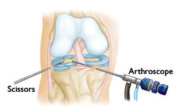
Knee arthroscopy
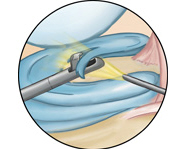
Close-up of meniscal repair
The surgeon will make 2 or 3 small incisions around your knee. Saltwater (saline) will be pumped into the knee to open up the space. A narrow tube with a tiny camera on the end will be inserted through one of the incisions. The camera is attached to a video monitor in the operating room. The surgeon looks at the monitor to see the inside of the knee and identify problems. Other medical instruments maybe introduced inside the knee through the other small incisions. The surgeon then repairs or removes the problem in the knee.
At the end of the surgery, the saline will be drained from the knee. The surgeon will close the incisions with sutures (stitches) and cover them with a soft bandage.
The range of procedures that can be performed at the time of standard knee arthroscopy include:-
- Meniscal resection / trimming / repair (as appropriate) for meniscal tears
- Abrasion chondroplasty (shaving of rough damaged cartilage for articular cartilage damage )
- Radiofrequency ablation chondroplasty (welding-over of cracks / fissures in the articular cartilage)
- Removal of cartilage or bone loose bodies
- Removal or repair of torn meniscal cartilage
- Reconstruction of a torn anterior cruciate ligament
- Trimming of torn pieces of articular cartilage
- Removal of inflamed synovial tissue
After procedure
Keep your leg elevated as much as possible for the first few days after surgery. Apply ice to relieve swelling and pain.
You should exercise your knee regularly for several weeks after surgery. This will restore motion and strengthen the muscles of your leg and knee.
Therapeutic exercise will play an important role in how well you recover. A formal physical therapy program may improve your final result.
Arthroscopy surgery can:
- Relieve pain.
- Improve joint stability.
- Repair tears and damage.
- Maximize quality of life.
- Optimize activities of daily living.